Project Description
Monitoring of 3D skin cell cultures
In Collaboration with the Radboud University Medical Centre.
At the Laboratory of Experimental Dermatology of the RadboudUMC, impedance measurements were implemented as a method to determine the barrier integrity of 3D skin cell cultures, shown in figure 1.
Skin cell cultures were derived from stem cells, which were extracted from skin tissue donated by three different patients. The impedance measurements were performed with the Artemis connected to 24 wells smart-lid to a NUNC transwell plate with inserts. First results show that with the Artemis the following cell culture variations could be monitored:
- barrier integrity of the cell cultures
- discrimination between the three patients
- applied damages to the skin culture
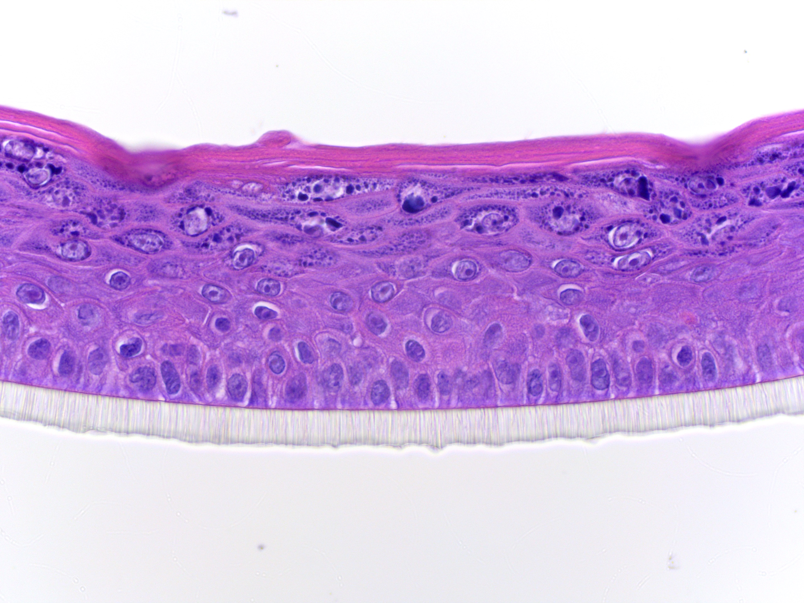
Figure 1 Microscopy image of the cross section of a 3D skin model showing the morphology. The culture was colored with hematoxylin and eosin. Material
- Skin cell cultures
- 24 wells NUNC transwell plate with inserts
- Locsense Artemis with smart-lid containing 24 gold plated electrodes.
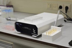
Figure 2a) Locsense Artemis with smartlid to a 24 wells NUNC transwell
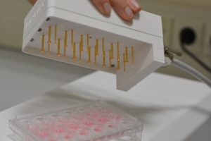
Figure 2b) Smart-lid containing 24 gold plated electrodes.
Results
From figure 3 it can be seen that an intact skin cell culture shows an overall higher impedance than a damaged skin cell culture. The shape of the curve is characteristic. Upon scratching of the cell culture with increasing severity, the measured impedance drops across the entire spectrum. An increase in permeability due to the skin damage is the cause of this observed drop in the impedance.
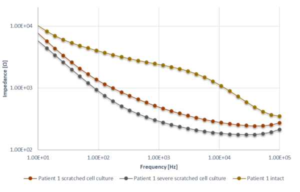
Figure 3) Impedance spectra for cell cultures from a single patient, Two skin samples were scratched and one sample was intact.
Figure 4 features the results of impedance measurements performed on nine samples derived from three patients. Three samples were obtained from each patient. As can be seen from the graph, the curves are clustered according to patient origin.
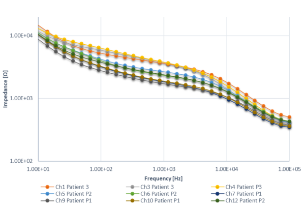
Figure 4) Overview of impedance measurements for skin cell cultures derived from three different patients. The measurements are grouped accordingly.